Cerebellum Fissure MRI: What It Is and What It Tells You.
Magnetic Resonance Imaging (MRI) is one of the most helpful and accurate tools to identify abnormalities in the brain. It helps determine things like a stroke, bleeds, and abnormalities in structure that indicate severe pathology. An area that often requires an MRI to diagnose issues is called the cerebellum. This complex area involves motor planning/execution, balance, eye movements, and thought. In this article, we’ll cover the importance of an MRI and why looking at the fissures within the tissue can help identify subtle issues that affect function and well-being.
What is the Cerebellum?
The cerebellum, which translates to “little brain” in Latin, is a vital structure that controls various essential functions. Located right behind the brain, the cerebellum is a small, densely packed ball of neural tissue containing more neurons than the entire brain combined. It needs this many connections because it fine-tunes every movement you make. It’s divided into parts that control different aspects of movement. For example, the midline cerebellum controls core muscles close to the spine, the middle portion controls joints like hips and shoulders, and the outer portion controls fingers and toes. The hemispheres are split into sections by a fissure, a common site for imaging.
What is a Cerebellum Fissure MRI?
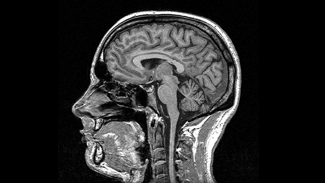
An MRI of a fissure is a scan to examine the integrity of the cerebellar lobes and the groves (fissures) that divide them. Each lobe has a specific function, and imaging the fissures can give you a more detailed understanding of pathology or functional damage to the cerebellar tissue. MRIs identify strokes, tumors, abscesses, or congenital abnormalities. They could be more helpful in identifying functional issues.
Why is a Cerebellum Fissure MRI Important?
Examining the fissures of the cerebellum is vital for several reasons. It is effective for evaluating the integrity of anatomical structures, like the tonsils and the division between lobes. It’s also helpful in assessing the progression of various neurological illnesses. Below is a list of abnormalities a cerebellar MRI can detect.
1. Cerebellar Atrophy
- Degenerative conditions can reduce the size and volume of the cerebellar lobes, which will show up as reduced depth of the cerebellar fissures. Other conditions, like chronic alcoholism, can shrink the cerebellar tissue, especially in the anterior lobe, and will present as shallow, poorly defined fissures.
2. Developmental Anomalies
- Certain congenital conditions, like a Dandy-Walker malformation, will show up on an MRI. You can identify this by the structure of the lobes and the definition of the cerebellar fissures.
3. Tumor Detection
- Tumors can alter the shape and morphology of the cerebellum. Astrocytomas and medulloblastomas show up as abnormalities in the shape and alignment of the fissures and surrounding tissue.
4. Inflammation and Infections
- Inflammatory conditions and autoimmune disorders often change the appearance of the cerebellar tissue and surrounding fissures.
5. Stroke Assessment
- MRI can easily detect Ischemic or hemorrhagic strokes. The restricted blood flow and possible bleeding within the tissue will cause a characteristic change in the shape and orientation of the fissures.
6. Vascular Malformations
- Arteriovenous malformations (AVMs) or cavernomas, clusters of abnormal blood vessels, can often be situated in or affect areas around the cerebellar fissures, which are visible on MRI scans.
7. Cysts
- If a cyst forms, it may change the appearance of the tissue or displace other tissue, causing a change in normal fissure anatomy on an MRI.
8. Degenerative Changes
- Multiple sclerosis and dementia can show plaque formations, which present as atrophy of the cerebellar cortices and abnormal boundaries of the fissure anatomy.
9. Hydrocephalus
- When the CSF pressure increases, the cerebellar fissures appear enlarged. This indicates compression of the cerebellar tissue, which is especially sensitive to compressive forces due to its orientation in the skull.
10. Traumatic Brain Injury (TBI)
- Due to its location, the cerebellum is susceptible to injury when it is pressed up against the skull during a head injury. Contusions to the tissue and changes in blood flow show up on an MRI.
11. Fissure Integrity
- In cases of compression or herniation, an MRI is helpful because it allows for a clear view of the fissure borders, which can quantify the degree to which it has been shifted or compressed.
How is a Cerebellum Fissure MRI Performed?
– Preparation: You’ll remove any metal objects because the machine uses a magnetic field to take the image. If the image is done with contrast, a contrast agent is used to improve the quality of the image.
– Scanning: While remaining still, the cylinder shaped MRI machine passes over the target tissue and uses the magnetic field to obtain the image. Your only job is to be as still as possible.
– Duration: A typical cerebellar MRI will take between 30-60 minutes from start to finish.
What Do Neurologists Look For in a Cerebellum Fissure MRI?
When interpreting an MRI of the cerebellum fissures, neurologists look for:
– Fissure definition: Small changes in normal fissure anatomy, such as defining borders and depth, can signal specific issues. Some of these are listed above.
– Atrophy or Enlargement: Atrophy can point to degenerative conditions, and swelling or enlargement often points to pressure changes or fluid accumulation.
– Lesions or Cysts: Abnormal tissue findings are explored further for masses that might indicate tumors or cysts.
– Blood Flow: Sometimes, the doctor looks for abnormalities to rule out ischemic stroke or hemorrhages.
Conclusion
An MRI is a valuable tool for ruling out different forms of disease. It is beneficial to analyze the cerebellar fissures because they provide a reliable baseline for normal anatomical boundaries. Alterations in fissure size, definition, depth, and shape can all point to different issues of varying severity. With the correct information, you and your healthcare provider can create a plan of action that suits your needs and gives you the best chance for long term success.
If you have questions about where to turn, click here to schedule a complimentary consultation with one of our doctors. We’ve treated hundreds of complex neurologic cases and can help piece together the missing pieces in your recovery.
*Note: The information provided in this article is for educational purposes only and does not constitute a doctor-patient relationship. Patients should consult their medical provider or primary care physician before trying any remedies or therapies at home.
